Home

Echivalent Curat câțiva pineal gland optic microscope opțiune plutitor Neîncetat

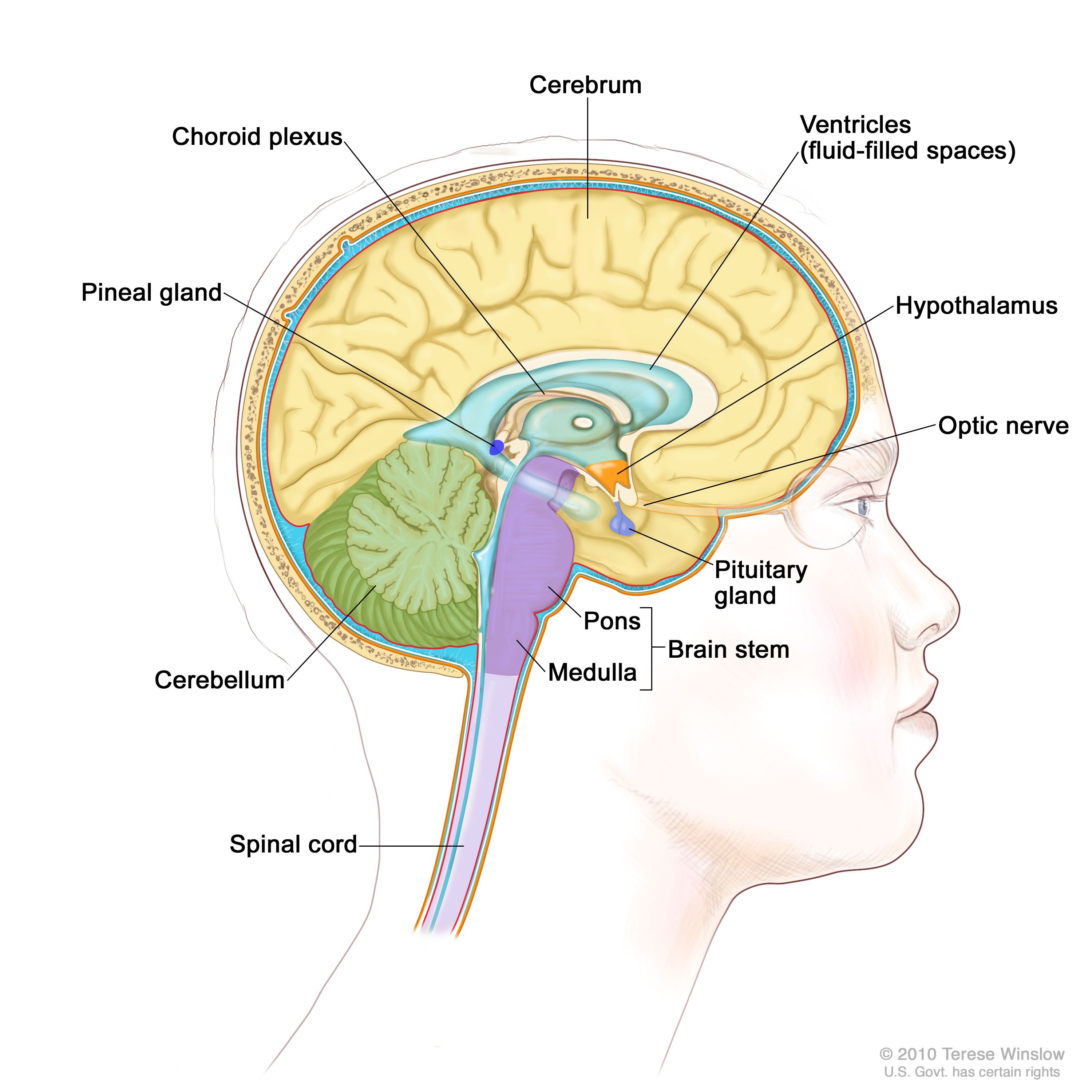
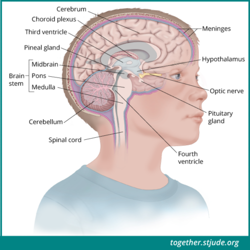
Pineoblastoma in Children and Teens - Together

Normal pineal gland producing melatonin and serotonin. Optical microscope  X100, Stock Photo, Picture And Rights Managed Image. Pic. VD7-2972552 |  agefotostock
Normal pineal gland producing melatonin and serotonin. Optical microscope X100, Stock Photo, Picture And Rights Managed Image. Pic. VD7-2972552 | agefotostock

Light Microscope of the Pineal at Higher Magnification

The pinealocytes of the human pineal gland: A light and electron microscopic  study. | Semantic Scholar
The pinealocytes of the human pineal gland: A light and electron microscopic study. | Semantic Scholar

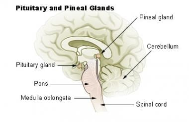
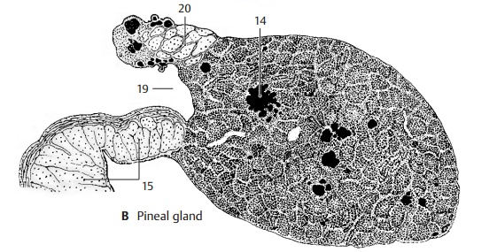
Pineal Gland Anatomy: Overview, Gross Anatomy, Microscopic Anatomy

Pineal Gland - Epithalamus

Does a pineal gland really exist? - Quora

Pineal gland - Wikipedia

Human Structure Virtual Microscopy

Pineal gland glial scars, light micrograph - Stock Image - C036/1344 -  Science Photo Library
Pineal gland glial scars, light micrograph - Stock Image - C036/1344 - Science Photo Library

Journal of Comparative Neurology | Systems Neuroscience Journal | Wiley  Online Library
Journal of Comparative Neurology | Systems Neuroscience Journal | Wiley Online Library

Anatomy Atlases: Atlas of Microscopic Anatomy: Section 1 - Cells

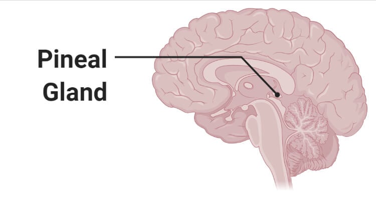
Pineal Gland- Definition, Structure, Hormones, Functions, Disorders

Investigation of the human pineal gland 3D organization by X-ray phase  contrast tomography - ScienceDirect
Investigation of the human pineal gland 3D organization by X-ray phase contrast tomography - ScienceDirect
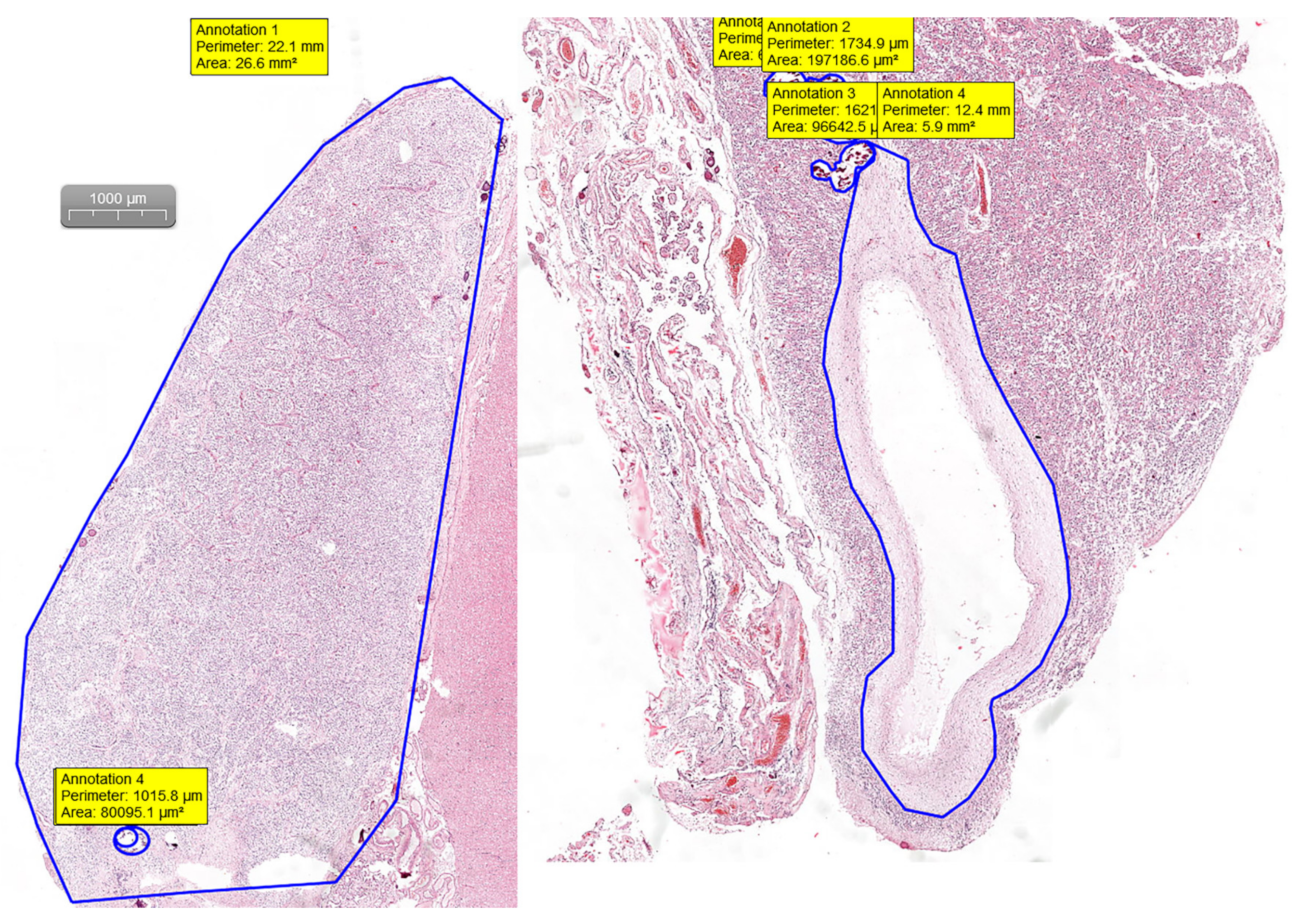
Medicina | Free Full-Text | Age-Related Changes of the Pineal Gland in  Humans: A Digital Anatomo-Histological Morphometric Study on Autopsy Cases  with Comparison to Predigital-Era Studies
Medicina | Free Full-Text | Age-Related Changes of the Pineal Gland in Humans: A Digital Anatomo-Histological Morphometric Study on Autopsy Cases with Comparison to Predigital-Era Studies

Pigmented Cells in the Pineal Gland of Female Viscacha (Lagostomus maximus  maximus): A Histochemical and Ultrastructural Study
Pigmented Cells in the Pineal Gland of Female Viscacha (Lagostomus maximus maximus): A Histochemical and Ultrastructural Study

The pinealocytes of the human pineal gland: A light and electron microscopic  study. | Semantic Scholar
The pinealocytes of the human pineal gland: A light and electron microscopic study. | Semantic Scholar
Pineal gland lobulation, light micrograph - Stock Image - C036/1341 -  Science Photo Library
Pineal gland lobulation, light micrograph - Stock Image - C036/1341 - Science Photo Library

Pinealocyte - Wikipedia

Pineal Gland | The Neurosurgical Atlas

Pineal region tumours (type of brain tumour in the pineal gland) |  Macmillan Cancer Support
Pineal region tumours (type of brain tumour in the pineal gland) | Macmillan Cancer Support

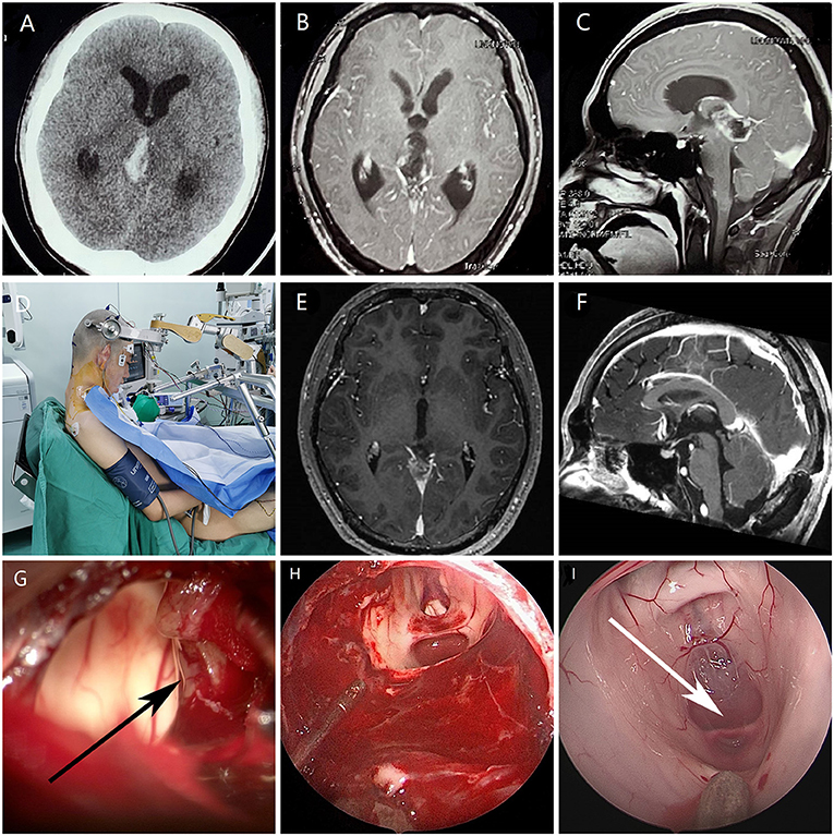
Frontiers | Endoscope-Assisted Microsurgery in Pediatric Cases With Pineal  Region Tumors: A Study of 18 Cases Series
Frontiers | Endoscope-Assisted Microsurgery in Pediatric Cases With Pineal Region Tumors: A Study of 18 Cases Series
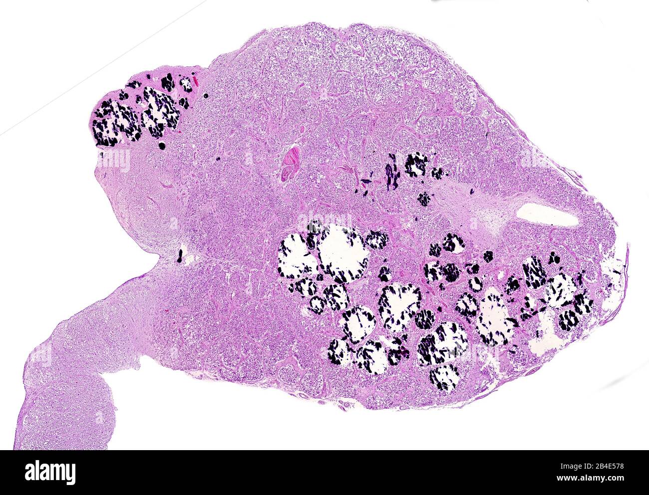
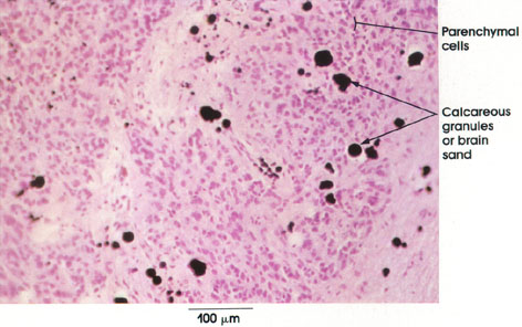
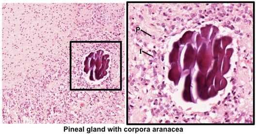
Sagittal section of a human pineal gland. Numerous calcareous concretions  can be seen, most of them broken by artefact. Light microscope micrograph  st Stock Photo - Alamy
Sagittal section of a human pineal gland. Numerous calcareous concretions can be seen, most of them broken by artefact. Light microscope micrograph st Stock Photo - Alamy
Human Structure Virtual Microscopy

Pituitary Tumors Treatment: Robert H. Lurie Comprehensive Cancer Center of  Northwestern University : Feinberg School of Medicine
Pituitary Tumors Treatment: Robert H. Lurie Comprehensive Cancer Center of Northwestern University : Feinberg School of Medicine

Growth patterns for acervuli in human pineal gland | Scientific Reports

Pineal gland, light micrograph - Stock Image - C036/1349 - Science Photo  Library
Pineal gland, light micrograph - Stock Image - C036/1349 - Science Photo Library

An ultrastructural study of the deep pineal gland of the Sprague Dawley rat  using transmission and serial block face scanning electron microscopy: cell  types, barriers, and innervation | SpringerLink
An ultrastructural study of the deep pineal gland of the Sprague Dawley rat using transmission and serial block face scanning electron microscopy: cell types, barriers, and innervation | SpringerLink